Biomedical engineers in the UK and the US have made software advancements that are enabling the monitoring of patients’ vital signs such as pulse rates through the analysis of standard video and webcam footage.
The breakthroughs could lead to less invasive diagnostic techniques and the emergence of more widespread tele-health applications, where patients are assessed by doctors and nurses virtually, from the comfort of their own homes.
The US researchers at Massachusetts Institute of Technology have developed an algorithm that can accurately measure the heart rates of people depicted in ordinary digital video by analysing imperceptibly small head movements that accompany the rush of blood caused by the heart’s contractions.
In tests, the algorithm gave pulse measurements that were consistently within a few beats per minute of those produced by electrocardiograms (ECGs). It was also able to provide useful estimates of the intervals between beats, a measurement that can be used to identify patients at risk of cardiac events.
A video-based pulse-measurement system could be useful for monitoring newborns or the elderly, whose sensitive skin could be damaged by frequent attachment and removal of ECG leads.
John Guttag, a professor of electrical engineering and computer science at Massachusetts Institute of Technology, said that, from a medical perspective, the technique’s long-term use could go far beyond just pulse measurement. “For instance,” he said, “an arterial obstruction could cause the blood to flow unevenly to the head. Could you use the same type of techniques to look for bilateral asymmetries? What would it mean if you had more motion on one side than the other?”
Similarly, Guttag said, the technique could, in principle, measure cardiac output, or the volume of blood pumped by the heart, which is used in the diagnosis of several types of heart disease. Indeed, he said, before the advent of the echocardiogram, cardiac output was estimated by measuring exactly the types of mechanical forces that the new algorithm registers.
In a technique called ballistocardiography, a heart patient would lie on a table with a low-friction suspension system; with every heartbeat, the table would move slightly, with a displacement corresponding to cardiac output.
“I think this should be viewed as proof of concept,” said Guttag. “It opens up a lot of potential flexibility.”
The algorithm works by combining several techniques common in the field of computer vision. First, it uses standard face recognition to distinguish the subject’s head from the rest of the image. Then it randomly selects 500 to 1,000 distinct points, clustered around the subject’s mouth and nose, whose movement it tracks from frame to frame.
Next, it filters out any frame-to-frame movements whose temporal frequency fall outside the range of a normal heartbeat — roughly 0.5 to 5Hz, or 30 to 300 cycles per minute. That eliminates movements that repeat at a lower frequency, such as those caused by regular breathing and gradual changes in posture.
Finally, using a technique called principal component analysis, the algorithm decomposes the resulting signal into several constituent signals, which represent aspects of the remaining movements that don’t appear to be correlated with each other. Of those signals, it selects the one that appears to be the most regular and that falls within the typical frequency band of the human pulse.
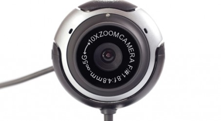
Meanwhile, Oxehealth, a spin-out company from Oxford University, has developed a technology that can use a webcam to track a patient’s pulse, breathing rate and oxygen saturation without the need for any other hardware.
The technology has been validated in a clinical study with patients in the Oxford Kidney Unit, showing that respiratory rate, pulse rate and oxygen saturation can all be monitored with a remote camera.
The measurement of the cardiovascular pulse using light reflectance or transmission has been employed clinically since the 1980s to monitor hospital patients using a finger probe sensor with in-built LEDs and a photodiode.
Cardiovascular signals can be remotely acquired without the use of body-worn sensors by measuring the light reflected from an individual’s face using a webcam. However, the interference caused by ambient, artificial light has hampered the application of the technology outside the research laboratory, rendering it impractical and unreliable for commercial application.
The Oxford invention includes novel algorithms that remove the effects of ambient light interference, allowing the technology to be used in everyday settings such as a patient’s home. Other algorithms are used to process the image recorded with the webcam and to extract from it the heart rate, respiratory rate and oxygen saturation.
Oxehealth’s academic founder, Professor Lionel Tarassenko, said the technology could be market-ready within two years.
